Home
Home Browse
 Close
Close Events
 Maps
Maps Email
Email Brightspace
 eCampus

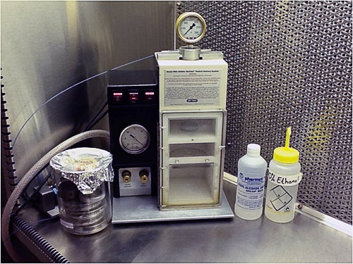
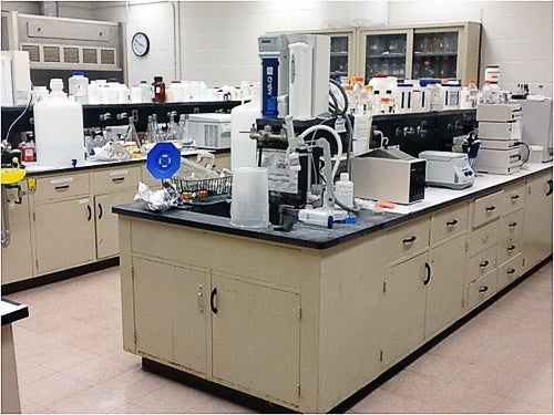
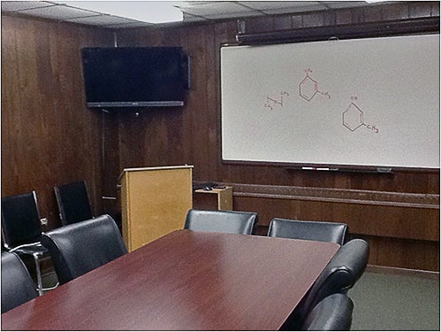
The PBL is comprised of research and teaching laboratories, a conference room, and faculty/staff office space. The PBL allows for training of faculty, staff and students (undergraduate and graduate) through participation in research programs requiring the routine production of transgenic materials. Various gene transfer capabilities are available including biolistics (PDS-1000/He Particle Delivery System, Bio-Rad), Agrobacterium-mediated gene transfer, microinjection, and electroporation (GenePulser II, Bio-Rad). A large suite of plant growth chambers and rooms are also housed in the PBL to support ongoing research. Administrative support and other routine office infrastructure (fax, copiers, slide duplicators, etc) are provided on site and through the main departmental office in the Center for Biotechnology and Life Sciences (CBLS) building on the URI campus. The PBL has been funded through foundation grants, as well as via industry, federal and state support.
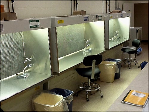
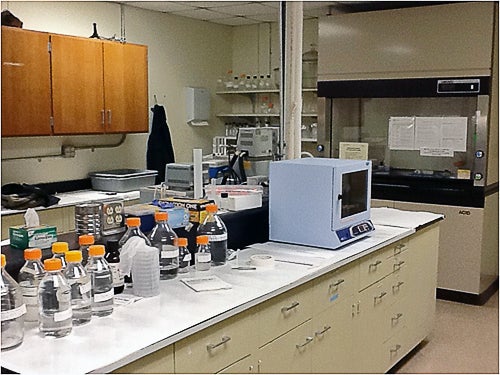
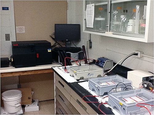
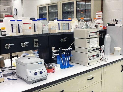
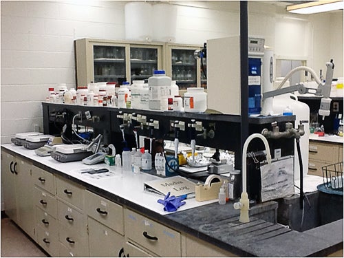
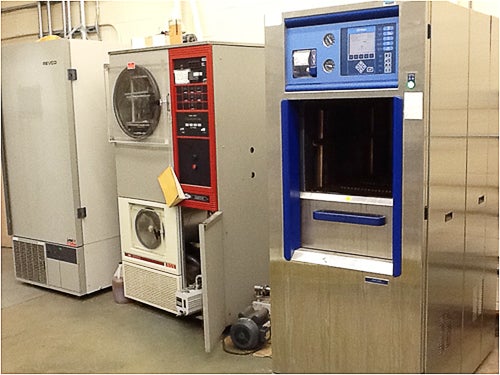
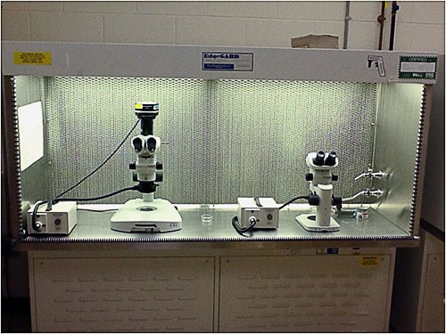
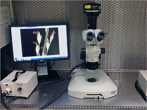
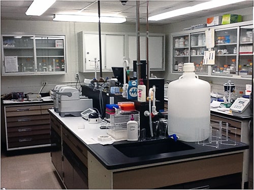
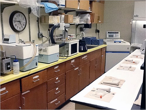
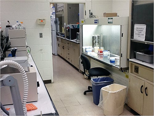
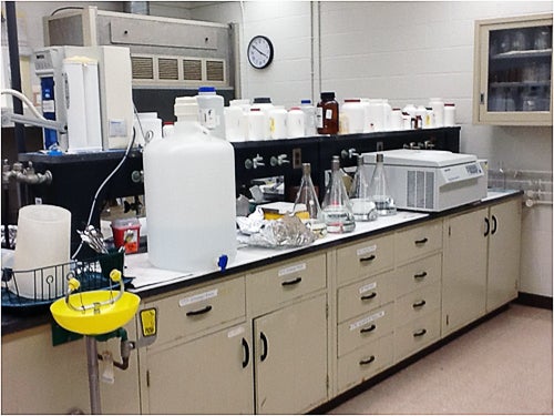
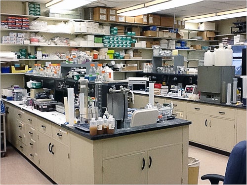
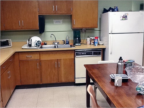
More than 4900 sq. ft. of laboratory space is equipped for plant transformation and molecular biology work. Various laboratories include a media lab (400 sq. ft.) with media preparation equipment, two 4°C refrigerators, a -20°C freezer, several water baths and pH meters. A centralized core laboratory (1200 sq. ft.) is used for both research and student demonstrations. This laboratory houses 6 laminar flow hoods equipped with stereo dissecting microscopes, an Olympus SZX7 stereo dissecting light microscope fitted with a CCD camera for image capture capability, and a PDS-1000/He Particle Delivery System (Bio Rad) for biolistic genetic transformation. Additional laboratory equipment includes multiple thermocyclers for PCR/RT-PCR (Eppendorf Mastercycler ProS) and qPCR (Applied Biosystems ViiA 7) work, a Kodak Pro Logic 112 Gel Documentation System, an Invitrogen Qubit 2.0 fluorometer and Thermo Fisher Scientific NanoDrop 2000 for nucleic acid quantification, as well as an ultracentrifuge, several microfuges and table top centrifuges. Vertical and horizontal electrophoresis units, power supplies, chemical fume hoods, incubators of all sizes, orbital shakers, multiple refrigerator/freezer units, a freeze dryer, two -80oC freezers, a speed-vac concentrator, and a vacuum oven are also available.
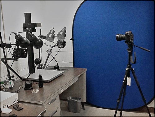
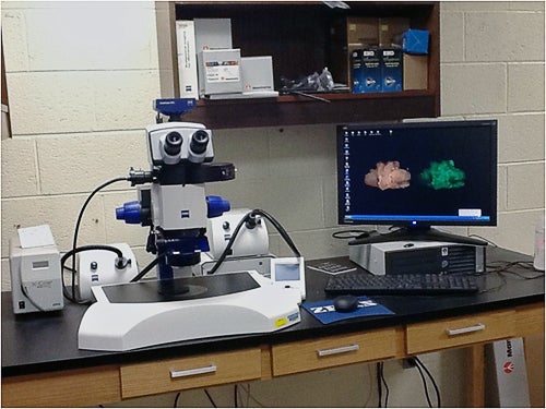
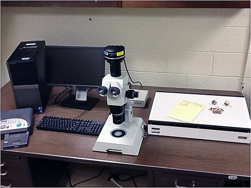
An array of photography and light microscopy equipment are on hand for publication quality image data capture including: two Olympus dissecting light microscopes fitted with CCD cameras; two Nikon high definition digital cameras with wide angle and macro lenses, light tables and tripods; a high definition digital video camera and blue screen; an image analysis system consisting of a Dell computer and either flatbed scanner input or video input; and, a Zeiss Discovery V2 GFP fluorescence microscope with an assortment of lenses.