1st Place | Photo Title: “Aquatic Oddball”
Michael Corso ’24, Aquaculture and Fisheries
Science
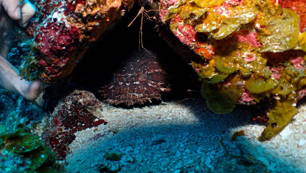
At 70 feet below the surface, a rare whitelined
toadfish peers out from the darkness to observe
a research dive group from URI. Corso captured
this photograph of a creature endemic to Belize’s
Barrier Reef system while on an aquaculture
and fisheries science J-term course in scientific
research diving. Corso says, “As an AFS major,
[I] focused on biological survey techniques and
underwater photography while collecting real
scientific data.” While the toadfish exemplifies the
extent of a reef’s ecosystem biodiversity, warming
seas and ocean acidification are chipping away
at the natural world’s biodiversity and weakening
reefs. “The highly specialized animals that rely
on these underwater jungles are being impacted
directly,” Corso says.
2nd Place | Photo Title: “Life is a Maze”
Janelle Mercer ’23, Marine Biology
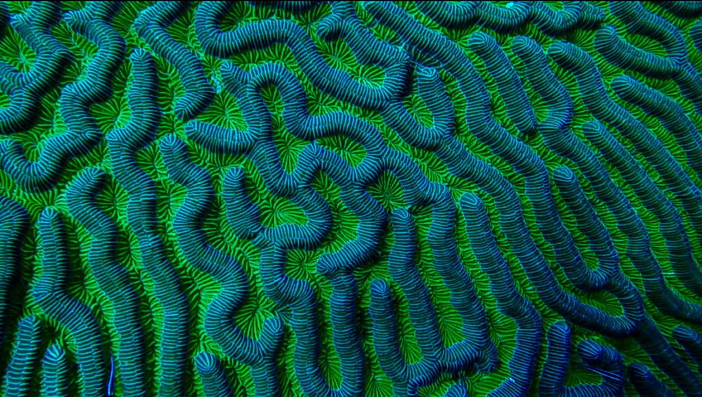
Mercer took this photo of maze coral off the coast of
St. George’s Caye, Belize, roughly 40 feet underwater,
during an underwater archaeology class with Anya
Hanson, Director, URI Diving Research and Safety
Program in Belize. Maze coral is a type of stony coral
with a photosynthetic dinoflagellate living within polyps
on the coral’s surface, providing coloration. The polyps
and their corallite walls have a unique twisting, maze
like formation. Mercer, who earned her AAUS Scientific
Research Diver certification on this trip, is preparing for
a career in marine biology and conservation.
3rd Place | Photo Title: “Got Nectar”
Julia Vieira, Graduate Student in Plant Sciences and Entomology
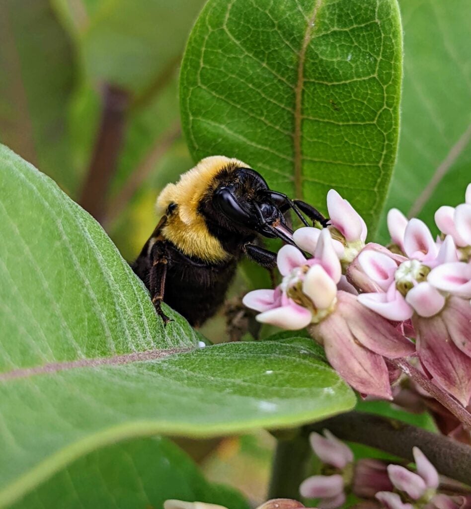
This macro photo shows a brown-belted bumble bee foraging for nectar
from common milkweed. The female worker takes a break to re-energize
by sucking up the delicious, carbohydrate-filled nectar within the milkweed
flower with her long proboscis (tongue). The bumble bee was visiting one
of the many milkweed plants within the acres of pollinator plantings on
URI’s East Farm. Vieira’s research primarily focuses on assessing bumble
bee visitation to various flower species to enhance Rhode Island bumble
bee conservation programs by improving floral recommendations for
pollinator plantings throughout the state.
Honorable Mention | Photo Title: “Crossing Under”
Olivia Mazzone ’23, Marine Biology
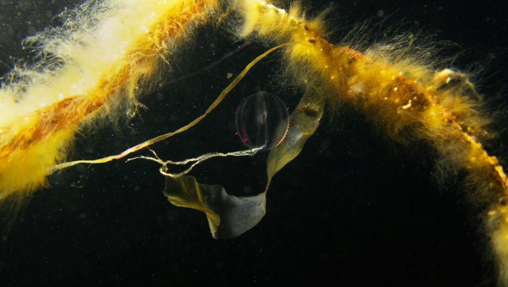
This photo depicts a comb jellyfish floating amongst
seaweed at dusk off the southeast corner of
Conanicut Island. “The day that I took this picture
was the first time I ever picked up an underwater
camera. It was an assignment for a class,” Mazzone
says. Her first attempts to photograph anything
underwater failed, she says, and she longed to
get out of the water and go home. “For whatever
reason instead of getting out of the water I lifted my
feet and let myself go completely. I became part of
the tide, and everything in my view became clear,”
she says—including this jellyfish, whom she now
considers “a dear friend.”
“There are things that I understand about the world that I can’t communicate in words,” Mazzone adds, but “to show people
life as I see it is an act of love.”
Honorable Mention | Photo Title: “Male Bombus Impatiens”
Gena Anika ’23, Wildlife and Conservation Biology
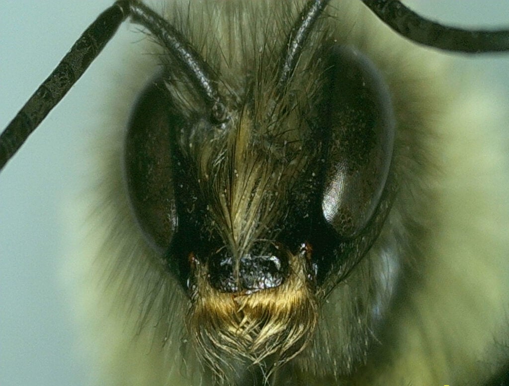
This photo is a close-up image of a Bombus impatiens
(Common Eastern Bumble Bee) face. The yellow patch of
hair on the bee’s face signifies it is male. There are pollen
granules present on the bee’s face and you can see the
hexagonal lenses (ommatidium) in the compound eyes.
Anika used a digital microscope to observe the bee closely
to help learn bee characteristics and to identify its species
and sex for the class BIO 338 Bees and Pollination.
Honorable Mention | Photo Title: “Fluorescence Microscopy of Neuromuscular Junctions”
Alyssa Madden ’23, Molecular Neuroscience
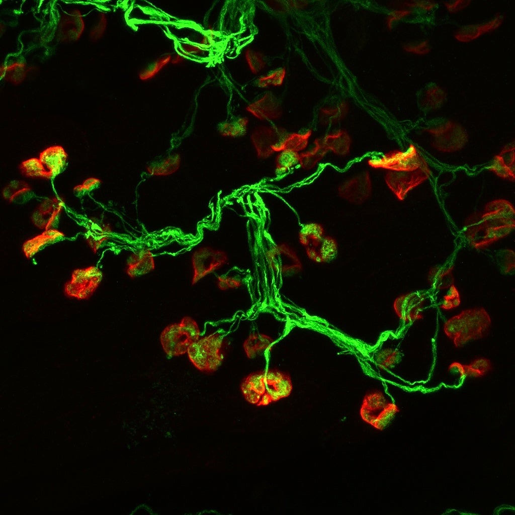
This fluorescence microscopy picture shows the neuromuscular
junctions in the calf muscle of a rabbit modeling cerebral palsy.
In the Manuel Lab, Madden is looking at the differences in
neuromuscular development in a rabbit model of cerebral palsy.
Using confocal microscopy, researchers can observe how the
structure of the neuromuscular junctions is affected by cerebral
palsy, in the hope of better understanding this disorder.