Atomic force microscopy (AFM) is a type of scanning probe microscopy that allows for imaging surfaces at the nanoscale level. It uses a tiny cantilever probe with a sharp tip to scan the surface of a sample, providing information about its topography, mechanical properties, and other characteristics.
AFM works by scanning the probe over the sample surface while measuring the force between the tip and the sample. This force information is then used to create a three-dimensional map of the sample surface with extremely high resolution, often on the order of nanometers or even angstroms.
AFM can be used to scan a wide variety of samples, including biological samples such as proteins, DNA, and cells, as well as materials like metals, semiconductors, and polymers. It can also be used to study surfaces in different environments, such as in air or in a liquid medium.
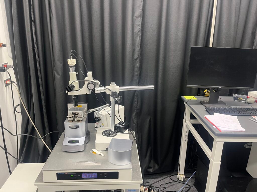
Instrument : Shimadzu Scanning Probe Microscope (SPM) 9700HT with 0.2 nm resolution in X ,Y directions and 0.01 nm in Z direction has the follwoing scanning modes: Contact, Dynamic, Phase, Lateral Force (LFM), Force Modulation Optional, Magnetic Force (MFM), Current, Surface Potential (KFM)
Location : Fascitelli Center for Advanced Engineering, Suite 060, 2 East Alumni Avenue, Kingston, RI 02881.
Instrument Manager : Matthew Cabral [Email: matthew.cabral@uri.edu]. Contact engimg@etal.uri.edu for instrument reservation.
Office: Fascitelli Center for Advanced Engineering, 2 East Alumni Avenue, Lower level suite 084, Kingston, RI 02881.
Contact engimg@etal.uri.edu for instrument reservation.
The use of the facility must be acknowledged in any publications by including the following text in the Acknowledgements section: “The AFM data was acquired at the RI Consortium for Nanoscience and Nanotechnology, a URI College of Engineering core facility.”