3D X-ray fluorescence (XRF) microscopy is a technique that uses X-rays to image and analyze the elemental composition of samples at high resolution. It provides three-dimensional images of the elemental distribution in a sample with micrometer to sub-micrometer resolution.
XRF works by using a tightly focused beam of X-rays to excite the atoms in a sample, causing them to emit characteristic fluorescent X-rays that can be detected and analyzed to determine the elemental composition of the sample. By scanning the sample with the X-ray beam and collecting the emitted X-rays, it is possible to create a three-dimensional map of the elemental distribution in the sample.
3D XRF microscopy can be used to scan a wide variety of samples, including materials such as metals, ceramics, semiconductors, and polymers, as well as biological samples such as tissues, cells, and organisms. It can be particularly useful for studying the distribution of trace elements, such as heavy metals or rare earth elements, in a sample.
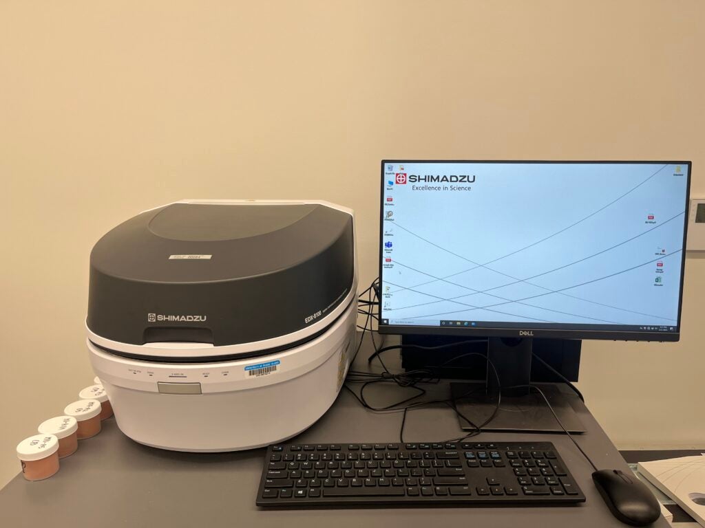
Instrument : Shimadzu Energy Dispersive X-ray Fluorescence Spectrometer EDX-8100 offers a high level of accuracy and speed in analyzing elements contained in various samples. For details and examples visit the Shimadzu webpage.
Location : Fascitelli Center for Advanced Engineering, Suite 051, 2 East Alumni Avenue, Kingston, RI 02881.
Instrument Manager : Matthew Cabral [Email: matthew.cabral@uri.edu]. Contact engimg@etal.uri.edu for instrument reservation.
Office: Fascitelli Center for Advanced Engineering, 2 East Alumni Avenue, Lower level suite 084, Kingston, RI 02881.
Contact engimg@etal.uri.edu for instrument reservation.
The use of the facility must be acknowledged in any publications by including the following text in the Acknowledgements section: “The XRF data was acquired at the RI Consortium for Nanoscience and Nanotechnology, a URI College of Engineering core facility partially funded by the National Science Foundation EPSCoR, Cooperative Agreement #OIA-1655221.”