EVOS Cell Imaging System 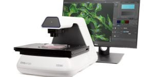
The EVOS M7000 microscope is a fully automated imaging system, incorporating both monochrome and color high-resolution CMOS cameras for the best of both fluorescent and colorimetric imaging. It includes up to 4 LED fluorescence light cubes and 5 parfocal objectives, focus, and exposure. Autofocus, Z-stack capability, image stitching and tiling, time-lapse imaging, and single-click multichannel capture. The M7000 can scan multi-well plates automatically and features speedy autofocus, image acquisition, and large data processing.
Sapphire Biomolecular Imager
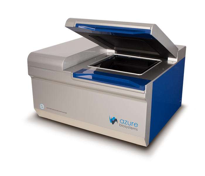 The Azure Sapphire Biomolecular Imager is a laser scanning system has four lasers (488, 520, 658 and 784 nm) giving a wide range of excitation. The instrument supports long and short wavelength Near IR fluorescence, red/green/blue imaging, phosphor imaging, and chemiluminescent imaging. Multiple detectors are used depending on the excitation wavelength and application for maximum sensitivity. It can accommodate gels up to 25cm by 25 cm.
IR Imager 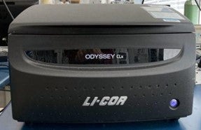
In the past, visible wavelength fluorescence technology has been widely used for protein and nucleic acid imaging applications. However, most visible systems have limited capabilities for imaging membranes due to strong background fluorescence at visible wavelengths. The Odyssey Infrared Imager eliminates these detection barriers by using near-infrared fluorophores. Low background fluorescence at infrared (IR) wavelengths provides a much higher signal-to-noise ratio than fluorescence detection at visible wavelengths. The sensitivity of the Odyssey system is comparable to chemiluminescence on film, but chemiluminescent substrates and film are not required. The Odyssey system typically uses two IR dyes for simultaneous two-color detection. Two-color detection adds many new detection capabilities. For example, two different antibodies can be used as probes on the same blot by using antibodies labeled with different IR dyes.
BioTek Cytation 5 Cell Imaging Multi-Mode Reader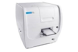
The BioTek Cytation 5 is a platform that combines automated microscopy and conventional microplate detection. It collects phenotypic and quantitative data in a single experiment.
Agilent Seahorse XFe96 Analyzer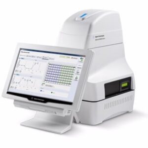
The Seahorse XFe96 Analyzers is a powerful tool used in real-time cell metabolic analysis. It measures the extracellular flux of key metabolites like oxygen and protons in live cells, enabling researchers to assess cellular bioenergetics, mitochondrial function, and cellular metabolism. It measures the oxygen consumption rate (OCR) and extracellular acidification rate (ECAR) of live cells in a 96-well plate format.
Digital Microscopes & Imaging System 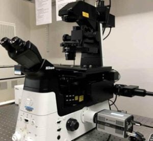
Nikon Eclipse Ti2 Inverted Confocal Microscope
The Nikon Eclipse Ti2 inverted confocal microscope features a high precision motorized-focus and vibration-free optical path changeover mechanism precisely controlled by high-precision Z-axis readout. This feature is perfect for research that requires comprehensive 3D information about the specimen. The C2Si point scanning confocal microscope system with its host of functions and various analytical capabilities is the perfect tool for the Nikon imaging system. Now fully controlled by NIS-Elements imaging software, the system includes four laser confocal imaging (405nm, 488nm, 561nm, and 640nm) and vastly expanded spectral capabilities with the ability to capture and unmix data acquired at any channel resolution across the entire detector bandwidth.
Nikon Eclipse E600 Upright Microscope 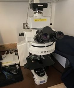 The Nikon Eclipse E600 upright microscope provides outstanding optical performance and versatility. This biological research microscope is equipped with Nikon’s CFI60 infinity optics for dramatically improved basic performance. Ideal for epi-fluorescence and other sophisticated microscopy, the E600 opens up new dimensions in advanced research applications.
Leica Microsystems Ivesta 3 Stereo Microscope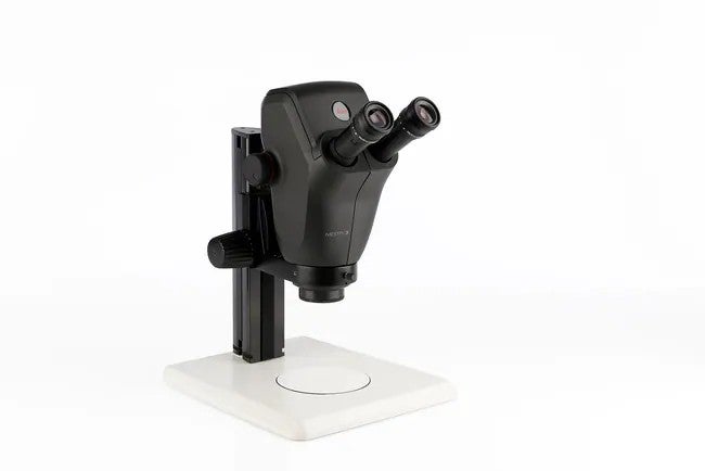
With this generation of Greenough microscopes operators will be able to reveal details faster as they spend less time having to adjust the microscope.
